Gong-hua Li ![]() ,
Chun-fang Lu
,
Chun-fang Lu
For correspondence:- Gong-hua Li Email: ligonghua133494@163.com Tel:+8657189972239
Received: 16 December 2016 Accepted: 3 July 2016 Published: 30 August 2016
Citation: Li G, Lu C. Effect of Salvia miltiorrhiza Bge extract on liver cirrhosis in rats. Trop J Pharm Res 2016; 15(8):1659-1562 doi: 10.4314/tjpr.v15i8.9
© 2016 The authors.
This is an Open Access article that uses a funding model which does not charge readers or their institutions for access and distributed under the terms of the Creative Commons Attribution License (http://creativecommons.org/licenses/by/4.0) and the Budapest Open Access Initiative (http://www.budapestopenaccessinitiative.org/read), which permit unrestricted use, distribution, and reproduction in any medium, provided the original work is properly credited..
Purpose: To explore the effects of Salvia miltiorrhiza Bge.extract(SMBE) on diethylnitrosamine(DEN)-induced liver cirrhosis in rats.
Methods: SMBE was obtained by extracting dried Salvia miltiorrhiza Bge. in water. Liver cirrhosis was induced in Wistar rats by injecting diethylnitrosamine in abdominal cavity once a week for 8 weeks. Concurrently, rats received either daily oral SMBE (SMBE group) or saline (control group). Clinical biochemical assessments, oxidative stress tests (malondialdehyde [MDA], superoxide dismutase [SOD]) were performed at 4 and 8 weeks after beginning DEN.
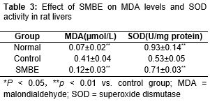
Results: Compared to the control group, both plasma alanine transaminase (ALT, 245.6 ± 8.5 U/L) and aspartate aminotransferase (AST, 205.7 ± 5.1 U/L) were significantly lower in SMBE group after 8 weeks (p < 0.01 for both ALT and AST). SMBE group exhibited significantly lower MDA (0.41 ± 0.04 µmol/L) levels and higher SOD(0.53 ± 0.05 U/mg protein) activity than control at 8 weeks after commencing DEN (p < 0.01 for both MDA and SOD).
Conclusion: SMBE has significant ameliorative effect on DEN-induced liver cirrhosis in rats. Anti-oxidant and anti-apoptotic effects of SMBE appear to be involved in these beneficial effects.
Introduction
Liver cirrhosis is a common pathological consequence of chronic liver disease, which is characterized by liver fibrosis, scar tissue and regenerative nodules, as well as leading to the destruction of the hepatic microstructure and liver dysfunction [1].
The structural changes include hepatic sinusoid capillarization, portal area and liver lobule fibrosis and alterations in microvascular structure. The dysfunction is manifested by the deficiency of liver function and portal hypertension. The main causes of liver fibrosis include hepatitis viruses, alcohol, drugs, toxins, schistosome, nonalcoholic steatohepatitis (NASH), cholestasis and autoimmune liver disease [2,3]. Their persistent insults on the liver activate hepatic stellate cells (HSCs) in the sinusoid, resulting in the imbalance of ECM metabolism. For example, ECM overproduction may cause over deposition in liver and hepatic structure remodeling. Liver fibrosis can progress into liver cirrhosis which causes further hepatocellular dysfunction and increases intrahepatic resistance to blood flow, leading to hepatic insufficiency and portal hypertension. Liver cirrhosis is the seventh leading cause of disease-related death in the United States [4]. Liver fibrosis was considered to be a passive and irreversible process due to the collapse of the hepatic parenchyma and its substitution with ECM components [5]. However, the reversibility of liver fibrosis has now been demonstrated both in patients and animal models [6].
Salvia miltiorrhiza Bge is a herb that possesses wide-reaching biological activities, including coronary artery dilation, improvement of myocardial ischemia, and modulation of the immune system, as well as antithrombotic, antioxidation, anti-aging, antihypoxic, antifatigue, anti-inflammatory, anti-hepatic fibrosis, antitumor, and analgesia effects[7-12]. As S. miltiorrhiza is used in traditional Chinese medicine to prevent liver cirrhosis[13], the current study was conducted to evaluate the anti-cirrhosis effects of the herb in rats. A diethylnitrosamine (DEN)-induced liver cirrhosis rat model was used, as this model closely resembles the cirrhosis observed in humans [5,6].
Methods
Plant material
Herbal samples of S. miltiorrhiza were collected from Bozhou City, Anhui Province, China, in May 2015. Taxonomic identification of the plant was performed by Professor HuChe of Zhejiang University, China. A voucher specimen of the herbarium (no. SMB 201505016) was deposited in the College of Pharmacy, Zhejiang University, China, for future reference. Aqueous S. miltiorrhiza extract (SMBE) was obtained by steeping dried S. miltiorrhiza in water at 60 °C for 1 h. The steeping process was performed three times. The sample was then dried in an oven, followed by freeze-drying to generate SMBE powder. One gram powder was obtained from approximately 2.2 g of crude Salvia miltiorrhiza Bge., which represents a yield of 45.5 %.
Animals
Male Wistar rats weighing 180 – 220 gwere provided by the Experimental Animal Center of Zhejiang Province (Certificate no. SYXK 2006 - 0008). The animals had free access to food and water and were permitted to acclimatize to their surroundings for at least 1 week before use. All experimental procedures involving the rats were approved by the Animal Care and Use Committee of Zhejiang University (approval ref no. 20100806) and conducted in compliance with the Directive 2010/63/EU on the handling of animals used for scientific purposes [14].
Animal group
The rats were randomly divided into 3 groups of 10 rats each: 1) normal group: negative controls, which were treated with neither DEN nor SMBE; 2) control group: control rats, which were injected with 60 mg DEN once per week for 8 weeks and administered oral saline solution once daily for 8 weeks, beginning just after DEN was started; and 3) SMBE group: experimental rats, which were injected with 60 mg DEN once per week for 8 weeks and administered 50 mg/kg oral SMBE once daily for 8 weeks, beginning just after DEN was started.
Blood chemistry
In the 4th and 8th week after beginning DEN injections, blood samples (0.5 mL) were obtained from each rat by retroorbital sinus puncture and collected in heparinized tubes. The blood was immediately processed for plasma extraction by centrifugation at 3500 g for 15 min. Plasma levels of alanine transaminase (ALT) and aspartate aminotransferase (AST) were measured by spectrophotometry, using commercially available kits (Nanjing Jiancheng Bioengineering Institute).
Determination of oxidative stress parameters
The rats were sacrificed by cervical dislocation, and their livers were excised, rapidly washed, and then homogenized in 10 volumes (v/w) of ice-cold saline solution. The homogenate was centrifuged at 3000 rpm for 10 min, and the supernatant was collected for subsequent use (as 10 % liver homogenate sample).
The level of malondialdehyde (MDA), a biomarker of lipid peroxidation, was determined spectrophotometrically by measuring thiobar-bituric acid reactive substances. One mL of 10 % trichloroacetic acid and 1 mL of 0.67 % thiobarbituric acid were added to 0.2 mL of the 10 % liver homogenate. The mixtures were incubated at 100 oC for 15 min. After cooling and centrifugation, the supernatant was aspirated and its absorbance (A) values at 532 nm and 600 nm were determined using water as blank. MDA concentration was calculated as in Eq 1.
MDA (μmol/L) = 6.45 (A532 – A600) ………. (1)
The superoxide dismutase (SOD) activity was measured spectrophotometrically, using commercially available SOD kit, A001-1 (Nanjing Jiancheng Bioengineering Institute).
Statistical analysis
Data are presented as mean ± standard deviation (SD). The results were analyzed using one-way analysis of variance followed by Tukey’s multiple comparison tests. SPSS 16.0 software for Windows was used for the statistical analyses. Differences with p < 0.05 were considered statistically significant.
Results
Effect of SMBE on ALT and AST levels in liver cirrhosis
During the weekly DEN injections, the body weight of the rats gradually decreased. After 4 weeks of DEN injections, the rats entered the hepatitis stage. DEN administration alone induced marked increases in plasma ALT and AST activity, whereas no increase in either enzyme was observed in the normal group (p < 0.01 for control vs. normal group at 4 weeks) (Tables 1 and 2). SMBE ameliorated this increase in ALT and AST: at 4 weeks both the ALT and AST were significantly lower in the control group than in the normal group (p < 0.01).
After the 8th week of DEN injections, liver cirrhosis was present in both the model and SMBE groups. Plasma concentrations of ALT and AST in the control group at 8 weeks were significantly lower than in the control group at 4 weeks (p < 0.05), but they remained higher than in the normal group (p < 0.05 for model vs. normal group at 8 weeks) (Tables 1 and 2). Plasma concentrations of ALT and AST were significantly lower in the SMBE group than in the control group at 8 weeks (p < 0.01).
Effect of SMBE on hepatic MDA and SOD in liver cirrhosis
Levels of MDA, a marker of oxidative stress, were significantly increased in DEN-treated control group rats (p < 0.01 for control vs. normal group) (). SMBE significantly reduced MDA formation in DEN-treated rat livers (p < 0.01 for SMBE vs. control group). Furthermore, SOD activity was significantly higher in the livers of rats treated with SMBE than in the livers of cirrhotic rats that did not receive SMBE (p < 0.01 for SMBE vs. control group).
Discussion
Liver fibrosis is characterized by overproduction and irregular deposition of extracellular matrix (ECM) in liver tissues, leading to the distortion of hepatic microstructure and liver dysfunction. Antifibrotic strategies against liver fibrosis include early intervention or control of etiologies, hepatic inflammation prevention and regulation of hepatic ECM metabolism and stellate cell activation. Viral hepatitis is the most important antecedent factor for liver fibrosis. Tremendous progress has been made in targeted antiviral treatment in recent years. Recent evidence showed that liver fibrosis could regress with effective antiviral treatment [15].
In recent years, many researchers have studied the anti-fibrosis properties of traditional Chinese medicine. The current study demonstrates that oral administration of SMBE exhibits the ameliorative effects on liver fibrosis and cirrhosis induced by DEN in rats. Potential mechanisms for these beneficial effects include the anti-necrosis, anti-apoptotic, and anti-inflammatory actions of SMBE, and/or SMBE-mediated reduction of activated hepatic stellate cells [16].
MDA, an end-product of lipid peroxidation, was significantly increased in our rat model of liver cirrhosis [17]. SMBE reduced MDA formation and also increased SOD activity in DEN-treated rats. SMBE likewise inhibited DEN-induced fibrosis, which appears to have been mediated by SMBE’s anti-oxidant activity. Furthermore, SMBE reduced plasma ALT and AST levels at both 4 and 8 weeks after beginning DEN injections, compared to DEN-treated rats that did not receive SMBE. All of these results suggest that SMBE attenuated apoptosis and oxidative stress in the fibrotic liver. Therefore, SMBE may be a potentially useful antifibrotic agent for future use in humans.
Conclusion
SMBE has significant ameliorative effects on DEN-induced liver cirrhosis in rats. The mechanism for inhibition of the fibrotic process appears to involve the anti-oxidant and anti-apoptoticproperties of SMBE.
References
Archives


News Updates